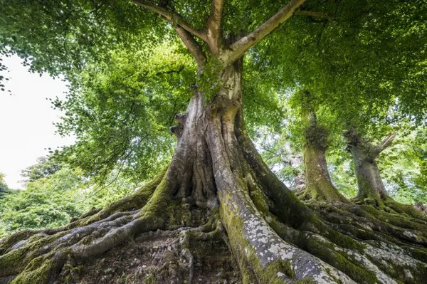
Plants, especially trees, are by far the tallest organisms on Earth. Height growth is made possible by a specialized vascular system. This system conducts water from the roots to the leaves with high efficiency, while simultaneously - thanks to extremely strong cell walls -providing stability. Through a combination of experiments and computer simulations, researchers from Wageningen University & Research in collaboration with colleagues in Germany and Australia have managed to study the formation of this system in more detail and to visualise it in a spectacular way. Their results were published today in Nature Communications and may in the future contribute to, among other things, the adaptation of plants and crops to climate change.
If one were to speak in fashion terms, one would say that plant cells in the vascular system make themselves a zebra-striped coat. They do not do this because it looks nice, but because it is a way for them to transport water from the roots to the leaves. They also don't want to be dependent on one single drinking straw, so to speak. If a straw is sealed at the bottom, it will squeeze together. The same would happen to the plant's vessels. Therefore, they use a 'zebra coat' instead.
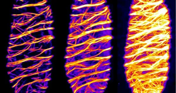
Xylem cells
Plants and trees take in water from the soil via the roots and transport it through a vascular system. This so-called xylem is a network of tubular cell wall containers that are formed by living xylem cells during plant growth. Before their death, the cells actively organize the deposition of an exceptionally strong wall - the so-called secondary wall - in unusual band and spiral patterns. After that, the cells dissolve and vacate their interior, and lignify the walls to further provide strength, resilience and waterproof to these structures. Due to this, xylem cells provide both an efficient water transportation system and the stability of plants and trees.
Cytoskeleton
The major load-bearing component of xylem walls is cellulose. To allow the cellulose to form these visually impressive band patterns, it needs the help of various proteins. These include so-called microtubules – small, tubular protein structures that are part of the cytoskeleton – which provide the molecular ‘tracks’ for the cell-wall-producing machinery. This machinery moves along the microtubules like an asphalt paver and continuously deposits wall material on the outside of the cells. The microtubules thus act like an instruction manual for cell wall synthesis. Despite much research into the general formation of cell walls, it has not yet been clear how the microtubule cytoskeleton is reorganized into such filigree patterns during secondary wall formation.
Gene switch
"One problem in elucidating these mechanisms is that the xylem cells are buried under many cell layers. As a result, they cannot be observed directly in the microscope. We use a genetic approach to make this process visible under the microscope," explains Kris van 't Klooster of the Laboratory of Plant Physiology.
The researchers used Arabidopsis thaliana (thale cress), an inconspicuous weed and model plant for research, and modified it in that way, that all its cells can be forced to form xylem and thus secondary cell walls. "For this purpose, we have equipped our plants with a 'gene switch'. This makes it possible to trigger the mechanism of xylem development from the outside in a targeted manner. By that, all cells in the plant become xylem cells, particularly the ones at the surface which are easy to study with high-resolution microscopy," Van 't Klooster describes. With this method, it is possible for the first time to observe xylem cells and their wall patterns as they develop.
Mapping the behaviour of microtubules
Using this new tool, the researchers describe which processes drive the rearrangement of the microtubules during xylem formation. They developed an automatic imaging method to collect data over a longer period of time and saw by means of detailed computer simulations that the microtubule bands and spirals form simultaneously over the entire cell surface and the resulting pattern is then further rearranged until an orderly distribution of the bands is achieved.
During this process, the microtubules in the gaps between the bands are continually broken down as they are growing in the bands. The re-arrangement into parallel, evenly spaced bands takes about one to two hours and is maintained for the remaining time.
The entire transformation process of a cell to become a proper xylem cell requires several days in total. Using their observations in plants and with the help of computer simulations, the team of scientists was also able to identify a protein complex, KATANIN, which turned out to be involved in the timely and orderly formation of secondary walls.
Adaptation to climate change
Based on these findings, fellow researcher Eva Deinum (Biometris) will work with PhD student Bas Jacobs to further investigate how exactly the patterns of secondary walls are formed in plants. This work is not only important for plant research, but could also contribute to the adaptation of plants to the future climate, as the survival of trees in a changing climate largely depends on the adaptive capacity of the xylem vessels.
The identification of proteins and associated genes that adapt the vascular system to environmental conditions could help identify or even genetically engineer more climate-resistant plant species.
 For more information:
Wageningen University & Research
www.wur.nl